Serial Endosymbiosis

Cellular evolution from microbes to eukaryotes.

Serial Endosymbiosis Theory (SET)

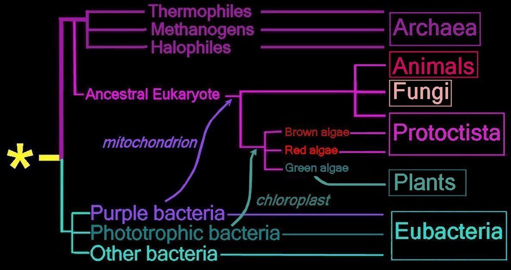
This widely accepted view of cellular evolution holds that the cellular evoluton of eukaryotes arose through the endosymbiotic union of engulfed bacteria with a precursor eukaryotic cell. The advent of oxygen releasing Cyanobacterial photosynthesis about 3 billion years ago (3 Ga) gradually converted the Earth's atmosphere from its primordial reducing state (timeline). Oxygen is toxic to organisms that lack the metabolic machinery to rapidly utilize oxygen, so symbiotic union with bacteria capable of oxidative metabolism would ensure cellular survival in environments with increased oxygen tension (pO2). This union ultimately resulted in mitochondrial organelles, while a second union with Cyanobacterial cells ultimately resulted in photosynthetic plastids (chloroplasts).

Constantin Mereschkowsky was the first to appreciate the significance of protists in early eukaryotic evolution. A.F.W. Schimper noted that chloroplasts in plant cells very much resembled cyanobacteria. The the ultimate theoretical model was provided by Lynn Margulis: the key step was the endosymbiosis of cyanobacteria within a phagotrophic eukaryotic host, a process she calls symbiogenesis. The symbiotic theory of mitochondrial origin is supported by the different nature of internal and external membranes in mitochondria.

In primary endosymbiosis, 1,000 genes were acquired by the nucleus from an incorporated cyanobacteria. A second round of gene transfer involved the engulfment of another plastid-containing eukaryote in secondary endosymbiosis. [S]

Prior to Lynn Margulis' conception of the Symbiotic Theory in the 1960's, biologists believed that the eukaryote's nuclear DNA coded for cellular organelles. When Margulis initially proposed the Symbiotic Theory, she predicted that organelles of prokaryotic origin would be coded for by their own DNA. In the 1980's, experimental evidence in support of Margulis’ prediction was found in the distinct prokaryotic-DNA of the mitochondria and chloroplasts of eukaryotic cells.

More detail:

“Much advance in evolution is due to the establishment of consortia between two organisms with entirely different genomes. Ecologists have barely begun to describe these interactions.” Ernst Mayr in foreword to Acquiring Genomes: A Theory of the Origins of Species, by Lynn Margulis and Dorion Sagan.

Biologist Lynn Margulis actively promoted endosymbiotic theory in the 1960s – The Endosymbiotic Theory of Eukaryote Evolution. Margulis published "Symbiosis in Cell Evolution" in 1981. Even though the essential idea had a lengthy history, mainstream biologists initially reacted to Margulis’ claims with incredulity and ridicule. On the basis of experimental evidence, Serial Endosymbiotic Theory (SET) is now almost universally accepted as the most plausible explanation for evolution of eukaryotes.

Serial Endosymbiotic Theory proposes that "symbiotic consortiums" of prokaryote cells were the ancestors of eukaryotic cells. In ecology, symbiosis indicates that two different organisms live in association with one another, and nature abounds with examples of ‘economic’ symbiotic relationships. Endosymbiosis is, in Margulis’ words, a ‘topological arrangement’, indicating that protracted symbiotic association generates an interdependent relationship in which the sum-of-the-parts becomes a new whole. As such, endosymbiosis generates evolutionary innovation where metabolic cooperation confers survival advantage.

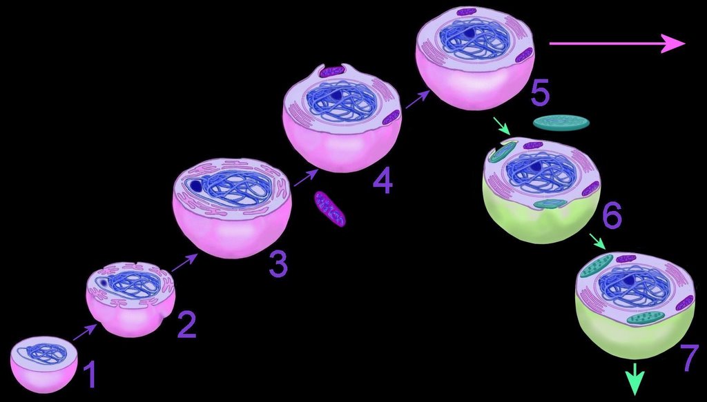
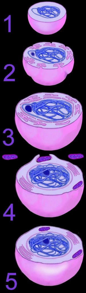
The mechanism of primary endosymbiosis is envisioned as phagocytosis of a bacterium (or bacteria) by another prokaryotic cell. (3-4) The phagocytozed bacteria survived upon nutrients from the host prokaryotic cell. Subsequently, both host and symbiotic bacterium reproduced co-independently such that subsequent generations of endosymbiotic neocytes would also contain the descendents of the originally ingested bacterium.

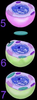
Ultimately, both the prokaryotic host and the bacteria endosymbionts developed an interdependence through endosymbiotic gene transfer, by which both entities lost their ability to function without the other (5). It is assumed that Cyanobacteria-generated oxygen in the early atmosphere necessitated endosymbiotic metabolic association between ingested aerobic bacteria and anaerobic host prokaryotes. The ingested bacteria ultimately performed oxidative metabolism necessary to the survival of the original host cell, which would otherwise have been poisoned by atmospheric oxygen. The former free-living aerobic bacteria assumed the role of mitochondria within its host cell (purple organelles within 5).

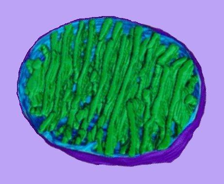
Similarly, serial ingestion of photosynthetic bacteria by endosymbiontic prokaryotes or eukaryotes (5-6) led to the evolution the ancestors of eukaryotic plants and photosynthetic protists (7). As the ingested photosynthetic bacteria adapted to the ingesting prokaryotic host cell, plastids, such as the chloroplast evolved (green organelle within 7). Primary plastids are found in Chlorophyta and plants, Rhodophyta, and Glaucocystophyta because their plastids are derived directly from a Cyanobacterium. All other lineages of plastids have arisen through secondary (or tertiary) endosymbiosis, in which a eukaryote already possessing plastids is engulfed by a second eukaryote. Considerable gene transfer has occurred among genomes and, at times, between organisms. A particularly complex history of plastid acquisition is found in eukaryotic crown group Alveolata. diagrams

[also]
References

Labels: , , , , , ,

| 0 Guide-Glossary

Experimental evidence for Endosymbiosis

Abundant evidence has been found in support of the concept of serial endosymbiosis:

1. Mitochondria and chloroplasts are similar in size and morphology to bacterial prokaryotic cells (though the mitochondria of some organisms are known to be morphologically variable).

2. Mitochondria and chloroplasts divide by binary fission, just as bacteria do, and not by mitosis as eukaryotes do. Both types of organelle have Fts proteins at their division plane.

3. Chemically distinct membrane systems:

The double membrane found in mitochondria and chloroplasts appears to be a relic of the absorption of the prokaryotic bacteria by the eukaryotic host cells. The inner membrane is of a different chemical composition (similar to that of prokaryotes) than the outer membrane of the organelle. Some organellar enzymes and inner membrane systems resemble prokaryotic inner membrane systems. The outer membrane is of similar composition to the plasma membrane of the eukaryote, as is the membrane of other cellular organelles such as the nuclear membrane, endoplasmic reticulum, and Golgi apparatus of eukaryotes (in support of the invagination hypothesis of their origin). Several primitive eukaryotic microbes, such as Giardia and Trichomonas possess a nuclear membrane yet have no mitochondria.

4. Mitochondria and chloroplasts have their own DNA and their ownribosomes:

The DNA of mitochondria and chloroplasts is different from that of the eukaryotic cell in which they are found. As Margulis predicted, both mitochondria and chloroplasts include DNA that is like that of prokaryotes – circular, not linear. Further, the DNA of mitochondria and chloroplasts, like that of the eubacteria, usually has neither introns nor histones. The first amino acid of mitochondrial and choloroplastic transcripts is equivalent to that of prokaryotes, and is different from that of eukaryotes.

Proteins encoded by mitochondrial DNA do not account for all of the mitochondrial proteins. The ingested prokaryotes are believed to have relinquished certain genes to the nuclei of their host cells, a process known as endosymbiotic gene transfer. For this reason, mitochondria and chloroplasts now depend on their hosts to synthesize most of their components.

The DNA of these mitochondrial and chloroplastic organelles evolves independently – and at a different rate – from the nuclear DNA of the eukaryotic cell. (Mitochondrial DNA is employed to trace evolutionary lines of human maternally-derived cells because virtually all DNA mtDNA is contributed by the oocyte, unlike nuclear DNA which derives from both parents, and unlike the Y-chromosome contributed solely by the human father.)

5. Mitochondria arise from preexisting mitochondria; chloroplastsarise from preexisting chloroplasts (they are not manufacturedthrough the direction of nuclear genes).

6. Organellar ribosomes are more similar in size to prokaryoticribosomes than to eukaryotic ribosomes:Mitochondria and chloroplasts produced their own ribosomes, which have 30S and 50S subunits, unlike the 40S and 60S subunits of the eukaryotic cells in which they occur.

7. Many antibiotics that kill or inhibit bacteria also inhibit proteinsynthesis of these organelles:

Antibiotics such as streptomycin block the synthesis of proteins in eubacteria, mitochondria, and chloroplasts, but not cytoplasmic protein synthesis in eukaryotes. Similarly, the antibiotic rifampicin infibits the RNA polymerase of eubacteria and mitochondria, but does not inhibit the RNA polymerase of the eukaryotic nucleus. Conversely inhibitors of eukaryotic protein synthesis, such as bacterially derived diphtheria toxin, do not affect protein synthesis within eubacteria, mitochondria, or chloroplasts.

8. Phylogenetic studies using comparative ribosomal RNA sequencing demonstrates that both mitochondria and plastids are related to the eubacteria. Phylogenetic analyses have clearly demonstrated that mitochondria and plastids derive from bacterial lines related to modern-day proteobacteria and cyanobacteria, respectively. Experimental observations confirm growth of bacterial endosymbionts in numerous organisms.

9. Microbiologist Kwang Jeon observed Legionella-like x-bacterial infection of strains of Amoeba proteus (xD) with which he was working. The infection killed many of the amoeba, but he raised the most hardy of the survivors. After many generations, the amoeba became dependent upon the bacterium, and endosymbiotic gene switching occurred. Free Full Text Article 2004 Detailed description xD amoeba experiments

References

| 0 Guide-Glossary

Secondary endosymbiosis

Secondary endosymbiosis and nucleomorph genome evolution: modified
The plastids (chloroplasts) of photosynthetic eukaryotes are the product of an ancient symbiosis between a heterotrophic eukaryote and a free-living Cyanobacterium. It is widely believed that this process, known as primary endosymbiosis, occurred only once and that all plastids descend from a single common ancestor. However, plastids have also moved laterally amongst unrelated eukaryotic cells by secondary endosymbiosis, a process that has occured multiple times and has given rise to a staggering array of photosynthetic organisms [diagram, see Archibald & Keeling 2002, Trends Genet. 18, 577- for review].

The Cryptomonads and Chlorarachniophytes are two microalgal lineages that are of particular interest with respect to secondary endosymbiosis. Unlike all other secondary plastid-containing algae, these organisms have retained the nucleus of the eukaryotic endosymbiont in a highly derived form called a nucleomorph. The nucleomorph genomes of Chorarachniophytes and Cryptomonads are very fast evolving and are the smallest eukaryotic genomes known, having transferred most of their genetic material to the nuclear genome of their respective host cells. Within the two groups, nucleomorph genome size varies considerably from lineage to lineage.

Continued evolutionary surprises among dinoflagellates -- Morden and Sherwood 99 (18): 11558 -- Proceedings of the National Academy of Sciences:"It is well established that chloroplasts in green and red algae are derived from a primary endosymbiotic event between a cyanobacterium and a eukaryotic organism 1 billion years ago (Fig. 1; refs. 1 and 2). Although these two groups account for many of the world's photosynthetic species, most other major taxonomic groups of photosynthetic organisms (stramenopiles including diatoms, phaeophytes, chrysophytes, and haptophytes) have plastids derived from a photosynthetic eukaryote implying a secondary endosymbiosis (1, 2). Still other groups, such as the dinoflagellates, have more complicated associations believed to be derived from tertiary endosymbioses involving the engulfment of a secondary endosymbiont. Each endosymbiotic event has characteristic structural changes associated with it, the most notable of which is the addition of two membranes surrounding the plastid (the inner representing the cell membrane of the engulfed organism and the outer representing the phagocytosis vacuole membrane) (2). Dinoflagellates, although believed to be tertiary endosymbionts, have only 3 membranes surrounding their plastids (1, 2), suggesting that the acquisition of too many membranes may be functionally unstable and can cause some to be lost. "
Clifford W. Morden, and Alison R. Sherwood Continued evolutionary surprises among dinoflagellates PNAS September 3, 2002 vol. 99 no. 18 11558-11560

Labels: , , , , , ,

| 0 Guide-Glossary

Mitochondrial origins

The eukaryotic mitochondrion (pl. mitochondria) is the 'power house of the cell. An outer membrane, similar in composition to the plasma membrane surrounds the organelle. The inner membrane is contiguous, at membrane junctions, with the inner membrane that forms the walls of cristae. The inner mitochondrial membrane contains more than 100 different polypeptides. The protein to phospolipid ratio is very high – more than 3:1 by weight, having about 1 protein for 15 phospholipids. The inner membrane is also rich in an unusual phospholipid, cardiolipin, which is usually characteristic of bacterial plasma membranes. This composition, along with other evidence, has led to the assumption that the inner membrane is derived from endosymbiotic prokaryotes.

Mitochondria are believed to have developed from an endosymbiotic union with alpha-proteobacteria, specifically the Rickettsiales. Phylogenetic analyses indicate that genome of R. prowazekii is more closely related to that of mitochondria than is any other microbe yet analyzed. Neither genome contains genes required for anaerobic glycolysis. R. prowazekii does contain a complete set of genes encoding components of the tricarboxylic acid cycle and the respiratory-chain complex, so ATP production in Rickettsia is the same as that in mitochondria. The genes from Rickettsia prowazekii encoding cytochrome b (cob) and cytochrome c oxidase subunit I (cox1) provide further phylogenetic evidence for a link with mitochondrial origins.

| 0 Guide-Glossary

History of ideas concerning endosymbiosis

1883 ~ AFW Schimper noted that the plastids of plant cells resembled free-living Cyanobacteria.
1905 ~ Mereschkowsky proposed a reticulated tree of endosymbiosis for the origin of algal plastids.
1920s ~ Ivan Wallin suggested a bacterial origin for mitochondria.
1959 ~ Stocking and Gifford discovered DNA in the plastids of Spirogyra, a green algae.
1960s ~ Lynn Margulis argued the case for endosymbiotic origins of mitochondria and plastids.
1970 ~ Margulis published her argument for the endosymbiotic origin of eukaryotes in The Origin of Eukaryotic Cells.
1977~ Carl Woese declared the case for prokaryotic endosymbiosis “clear cut” and “proven”. Other biologists subsequently declared the endosymbiotic theory demonstrated beyond a reasonable doubt.
1981 ~ In Symbiosis in Cell Evolution, Margulis argued that eukaryotic cells originated as communities of interacting entities. She extended the argument to including endosymbiotic incorporation of spirochaetes that developed into eukaryotic undulopodia -- flagella and cilia. (This proposal has not gained wide acceptance because flagella lack DNA and do not show ultrastructural similarities to prokaryotes.)

References

| 0 Guide-Glossary

Endosymbiotic transfers

Right - click to enlarge image: Proposed endosymbiotic transfer events between the three Domains and the six Kingdoms of Life. Both the Eubacteria and Archaea are prokaryotes, while animals, fungi, plants, and protists are eukaryotes.

The yellow asterisk * indicates the last universal common ancestor (LUCA), or universal cenancestor, which is hypothesized as being at the ancestral root of all living organisms. Not the earliest or simplest living organism, and not necessarily the sole example of its type, this organism possessed the genetic material that diverged (about 3.5 Ga) into all current living organisms. This diagram is not a cladogram, so branches do not indicate evolutionary timelines.

A number of terms are employed to refer to the universal cenancestor – last universal ancestor (LUA), last common ancestor (LCA), or last universal common ancestor (LUCA).

Woese and Fox proposed the Three Domain system: Eubacteria, Archaea, and Eukaryotes.

The Five Kingdom system was proposed in 1969: Monera (prokaryotes), Protista, Plantae, Fungi, Animalia. Discovery of the Archaea added the sixth kingdom.

History of taxonomic concepts:
Linnaeus, 1735 – 2 Kingdoms – Animalia, Vegetabilia
Haeckel, 1866 – 3 Kingdoms – Protista, Plantae, Animalia. Image Haeckel's tree of life.
Chatton, 1937 – 2 Empires – Prokaryota, Eukaryota
Copeland, 1956 – 4 Kingdoms – Monera, Protoctista, Plantae, Animalia
Whittaker, 1969 – Monera, Fungi, Protista, Plantae, Animalia
Woese et al, 1977 – 6 Kingdom – Eubacteria, Archaea, Protista, Fungi, Plantae, Animalia
Woese and Fox, 1999 – 3 Domain system: Eubacteria, Archaea, and Eukaryotes

References

| 0 Guide-Glossary

Endosymbiotic Gene Transfer

Proteins encoded by mitochondrial DNA do not account for all of the proteins found in mitochondria. Endosymbiotic prokaryotes are believed to have relinquished certain genes to the nuclei of their host cells in a process known as endosymbiotic gene transfer. For this reason, mitochondria and chloroplasts now depend on their host's DNA to direct synthesis of most of their components.

Microbiologist Kwang Jeon has demonstrated endosymbiotic gene transfer within endosymbiotic strains of Amoeba proteus (xD) with which he has worked since the 1970s. (J Eukaryot Microbiol 1997 Sep-Oct;44(5):412-9 Evidence for symbiont-induced alteration of a host's gene expression: irreversible loss of SAM synthetase from Amoeba proteus. Choi JY, Lee TW, Jeon KW, Ahn TI)
Free Full Text Article 2004 : Detailed description xD amoeba experiments

References

| 0 Guide-Glossary

Horizontal Gene Transfer

Horizontal gene transfer - gene swapping - has blurred the evolutionary relationships (phylogeny) of prokaryotes (image), and continues to provide a mechanism for the sharing of antibiotic resistance between bacteria.

(see The net of life: Reconstructing the microbial phylogenetic networkV. Kunin, L. Goldovsky, N. Darzentas, and C. A. OuzounisGenome Res. 1 July 2005. pdf)

Three mechanisms of horizontal (lateral) gene transfer are recognized: direct bacterial conjugation, bacteriophage mediated transduction between bacteria, and bacterial transformation by uptake of DNA fragments.

A major form of vertical gene transfer followed serial endosymbiotic events, in which ingested purple bacteria and Cyanobacteria became eukaryotic mitochondria and chloroplasts respectively. The ingested prokaryotes are believed to have relinquished certain genes to the nuclei of their host cells, a process known as endosymbiotic gene transfer.

Horizontal genomics is a new field in prokaryotic biology that examines DNA sequences in prokaryotic chromosomes that appear to have originated from other prokaryotes or eukaryotes. The prokaryotic mobile gene pool is referred to as the 'mobilome'. Various agents agents occur in all prokaryotes and effect DNA movement: plasmids, bacteriophages and transposons.

| 0 Guide-Glossary

Diagrams of proposed mechanism of SET

Simplified diagrams of serial phagocytosis of purple and photosynthetic bacteria, leading to primary* endosymbiotic accommodations. (adapted from)

1. ancestral prokaryote, undergoes

2. infoldings of plasma membrane, which permits


3. development of nuclear membrane and endomembranous system


4. engulfment of aerobic heterotrophic prokaryotes, generates


5. ancestral heterotrophic eukaryote with mitochondria.


~~~

Next, acccording to SET,

an ancestral heterotropic eukaryote (5)


undergoes
6. serial engulfment of photosynthetic prokaryotes, which generates


7. ancestral photosynthetic eukaryote with plastids.


Schematic in FFT Article Protist Images: Endosymbiosis and Parasitism Hatena

*Secondary endosymbiosis is engulfment by a eukaryotic cell of another eukaryote that already possesses endosymbiotic organelles derived from primary endosymbiosis. Similarly, acquisitions may be tertiary. The eukaryotic crown group Alveolata has a particularly complex history of plastid acquisition.


More images: Site with good diagrams tems & drop-down menu of endosymbiosis and chloroplasts : diagram of plastid diversity : diagram ~ endosymbiotic formation of algal groups :

References

| 0 Guide-Glossary

. . . endosymbiotic union began 10/06/06